[email protected] +92-320-6688665 Defence Road, Sialkot 51310, Pakistan
ISO 13485:2016 · CE Marked · FDA Registered Exporting to 45+ Countries
0%

Choosing the right Tumescent Infiltration Cannulas can influence comfort, control, and procedural efficiency. The choice is not merely about length or appearance. Experienced clinicians consider cannula design, tissue characteristics, infiltration volume, and the planned treatment area. A device that feels balanced in the hand may reduce unnecessary wrist movement during a long procedure.

Small details matter. The side ports should support even fluid distribution without excessive resistance. The connector must fit securely with the selected tubing and syringe. Cannula length should reach the target area without forcing awkward angles. Gauge selection also requires care, because flow rate and patient comfort can change with diameter. These decisions should follow the manufacturer’s instructions, institutional protocols, and applicable medical-device requirements.

Real-world evaluation is essential. A cannula may perform well in one patient but feel less suitable in fibrous or delicate tissue. That limitation deserves attention. Clinicians should check packaging integrity, sterility, and component compatibility before use. They should also monitor patient response throughout infiltration and adjust technique when appropriate. No single design is ideal for every situation.

This guide presents seven practical tips for comparing Tumescent Infiltration Cannulas. It focuses on handling, port configuration, sizing, materials, safety features, and reliable supplier information. The recommendations are educational, not a substitute for clinical training or professional judgment. Even familiar equipment deserves a careful review. Small choices can affect the entire procedure.
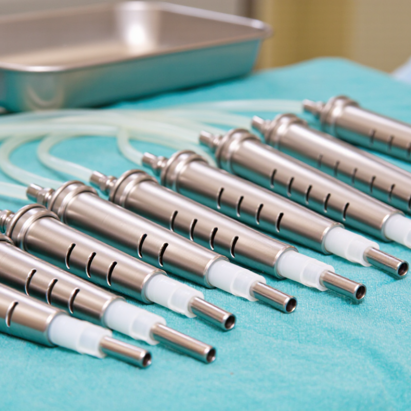
7 Tips for Choosing Tumescent Infiltration Cannulas

Define Cannula Requirements by Length, Gauge, Tip Geometry, and Flow

7 Tips for Choosing Tumescent Infiltration Cannulas

Define Cannula Requirements by Length, Gauge, Tip Geometry, and Flow

Procedure volume makes cannula selection more than a purchasing detail. The American Society of Plastic Surgeons’ 2023 Procedural Statistics reported more than 249,000 liposuction procedures in the United States. The 2023 ISAPS Global Survey recorded approximately 1.9 million procedures worldwide. These figures show demand, not a universal device specification. Choose length from the treatment field. Shorter cannulas improve control in compact areas. Longer shafts may reduce repeated repositioning across broad fields. Measure working distance, not the clinician’s reach.

Gauge should match intended flow and tissue resistance. Smaller internal diameters may restrict delivery. Larger diameters can increase fluid movement and handling demands. Verify actual inner diameter, not gauge alone. Tip geometry also changes dispersion. Multi-port tips may spread fluid broadly, while single openings create a more directed stream. Inspect port count, angle, and edge finish carefully. Sharp edges matter.

Request validated flow data at stated pressure, viscosity, and temperature. The same cannula may perform differently with saline and more viscous solutions. Do not guess. Review biocompatibility records, sterilization status, and instructions for use. Confirm connector compatibility before the procedure. A selection worksheet should record length, gauge, tip design, and measured flow. My own criteria would still need revision when tissue resistance or operator technique changes.

7 Tips for Choosing Tumescent Infiltration Cannulas

Define cannula requirements by length, gauge, tip geometry, and flow.

How to read the chart: The relative hydraulic flow index is calculated from the nominal gauge diameter using the Hagen–Poiseuille relationship, where flow capacity changes approximately with the fourth power of internal radius. It compares gauges at the same cannula length and assumes the same fluid and pressure. Actual flow varies with lumen design, fluid viscosity, cannula length, connector resistance, and tip configuration.

  1. Choose length by access depth: Select a length that reaches the intended treatment plane without excessive external leverage.
  2. Match gauge to fluid viscosity: Larger-bore cannulas generally provide greater potential flow, especially with more viscous solutions.
  3. Account for cannula length: For the same gauge, a longer lumen creates greater resistance than a shorter lumen.
  4. Evaluate tip geometry: Blunt, rounded, or atraumatic tips are commonly selected when tissue protection and controlled infiltration are priorities.
  5. Review outlet design: Single-hole, multi-hole, and side-port configurations distribute fluid differently and can affect coverage and injection pressure.
  6. Check compatibility: Confirm that the cannula, syringe, tubing, luer connection, and intended solution are compatible and securely connected.
  7. Prioritize controlled delivery: Select a configuration that supports predictable flow, visibility, maneuverability, and the operator’s established clinical protocol.

Compare 16–18G Cannulas for Controlled Flow and Reduced Tissue Trauma

7 Tips for Choosing Tumescent Infiltration Cannulas

Choosing between 16G and 18G cannulas starts with the fluid and the treatment area. A 16G cannula usually supports faster delivery with less resistance. An 18G cannula offers slower, more measured infiltration. Flow depends on length, side-port design, fluid viscosity, and applied pressure. Gauge alone does not tell the whole story.

Match the cannula to the tissue plane and the clinician’s control. Blunt tips and well-positioned openings may help distribute fluid evenly and limit concentrated pressure. A smaller 18G profile can reduce puncture size, but it may require more pushing force. Excessive force is a warning sign. Stop and reassess. Check sterility, connector security, and compatibility with the selected tubing or pump. These details are easy to overlook.

In practice, I compare flow before patient use. I watch for pulsing, leakage, and unexpected resistance. A 16G cannula may save time, yet faster flow can reduce tactile feedback. An 18G cannula can feel more controlled, although it may be inefficient for larger volumes. My own preference has changed with tissue density and fluid temperature. That is worth admitting. Document the cannula, flow setting, and observed response, then review the result against local protocols and current clinical guidance.

Match Delivery to 0.05–0.1% Lidocaine and 1:1,000,000 Epinephrine

Choosing tumescent infiltration cannulas starts with the solution, not the catalog. At 0.05–0.1% lidocaine, the concentration equals 0.5–1 mg/mL. Epinephrine at 1:1,000,000 equals 1 microgram/mL. The cannula should distribute this dilute mixture evenly, without excessive force. A multiport cannula may suit broad, superficial coverage. A smaller single-port design can offer more controlled placement. Match the outlet pattern to the intended tissue plane.

Check the internal diameter, length, tip design, and dead space. Narrow tubing may increase resistance and tempt operators to push harder. That is a warning sign. Confirm connector security, priming, and flow consistency before infiltration. Calculate total lidocaine exposure carefully, including all sources. The FDA Lidocaine Hydrochloride Injection Prescribing Information emphasizes dose limits, patient factors, and systemic toxicity monitoring. Concentration alone is not a safety plan.

The American Society of Plastic Surgeons reported 249,528 liposuction procedures in its 2023 Procedural Statistics, showing why repeatable infiltration practices matter. Choose a cannula that supports measured delivery, clear visualization, and gradual movement. Monitor vital signs and early neurologic symptoms throughout the procedure. I would not pretend that one cannula fits every patient. Tissue thickness, fibrosis, body region, and operator technique change the result. Review the setup with trained clinical staff, and follow current local regulations and institutional protocols.

Set Safety Limits: Published Tumescent Doses Reach 35–55 mg/kg Lidocaine

7 Tips for Choosing Tumescent Infiltration Cannulas

Published tumescent protocols report lidocaine doses of 35–55 mg/kg. These figures are not automatic safety targets. Dose selection depends on concentration, absorption, treatment area, liver function, body composition, and monitoring capacity. In practice, clinicians should calculate the total lidocaine amount before infiltration, including every anesthetic source. Keep a written record. Small calculation errors can become serious.

Tip 1: Choose a cannula that delivers controlled flow with minimal tissue trauma.
Tip 2: Confirm its length and diameter match the planned treatment layer. A short cannula may encourage repeated passes.
Tip 3: Use clear labeling for concentration and volume. The numbers must be visible.
Tip 4: Recheck the patient’s medicines, allergies, cardiac history, and hepatic risks before proceeding.

The 35–55 mg/kg range comes from published experience, but evidence and protocols differ. A higher reported dose should never replace individualized assessment. Lidocaine toxicity may appear later, especially after large-volume infiltration. Staff should recognize early symptoms, maintain appropriate observation, and follow an established emergency protocol.

Tip 5: Select equipment that supports steady, predictable infiltration.
Tip 6: Stop when tissue resistance, pain, or patient symptoms change unexpectedly.
Tip 7: Document the cannula, concentration, calculated dose, timing, and monitoring findings.

No single cannula makes an aggressive dose safe. That is an uncomfortable point. Better decisions come from conservative calculations, trained supervision, and willingness to revise the plan.

Verify Sterility, Luer-Lock Fit, and Performance Data Before Selection

7 Tips for Choosing Tumescent Infiltration Cannulas

Sterility is the first checkpoint, not a paperwork detail. Choose individually packaged, sterile, single-use cannulas with intact seals and clear lot information. The WHO Patient Safety Fact File reports that about one in ten patients experiences harm during healthcare, with more than half considered preventable. That figure is not cannula-specific, but it shows why basic contamination controls matter. Verify the sterilization method, expiry date, and storage conditions. Keep records.

Tip 1: Inspect every package before use.

Tip 2: Confirm the connector’s Luer-lock fit with the intended syringe and tubing. A secure connection should seat smoothly, resist accidental separation, and show no leakage during a controlled check. ISO 80369-7 specifies design requirements for small-bore Luer connectors, but compliance alone does not prove clinical performance. Ask for current conformity evidence.

Tip 3: Request performance data, including flow rate, pressure range, dead space, and leakage testing. Compare results using the same fluid, temperature, and test setup. Marketing tables can hide important differences.

Tip 4: Review needle orifice design and cannula flexibility for consistent delivery across the planned treatment area.

Tip 5: Confirm clear labeling and traceability.

Tip 6: Check staff handling instructions.

Tip 7: Record failures, even minor ones.

One weak connection can change the whole procedure. My own checklist once focused too heavily on sterility and missed flow consistency; that was a useful correction.

Only validated, documented evidence should support selection.

FAQS

How should cannula length be selected?

Measure the working distance across the treatment field. Shorter cannulas offer better control in compact areas. Longer shafts may reduce repositioning. Do not choose length based only on clinician reach.

Does gauge alone determine cannula performance?

No. Check the actual inner diameter, intended flow, and tissue resistance. A smaller opening may restrict delivery. A larger opening can increase fluid movement and handling demands.

How does tip geometry affect infiltration?

Multi-port tips can distribute fluid broadly. Single openings create a more directed stream. Inspect port number, angle, and edge finish. Sharp edges deserve attention.

What flow information should be requested?

Request validated flow data at stated pressure, viscosity, and temperature. Saline and thicker solutions may behave differently. Do not guess from gauge alone.

What safety checks should occur before infiltration?

Calculate the total anesthetic amount from every source. Review medicines, allergies, cardiac history, and liver risks. Record concentration and volume clearly. The numbers must be visible.

Are published lidocaine ranges automatic safety limits?

No. Published ranges of 35–55 mg/kg are not universal targets. Dose decisions depend on absorption, treatment area, body composition, and monitoring capacity. Higher is not automatically better.

What symptoms or changes should make the clinician stop?

Stop when resistance, pain, or patient symptoms change unexpectedly. Lidocaine toxicity can appear later. Staff need observation procedures and an established emergency response plan.

What should the procedure record include?

Document cannula length, gauge, tip design, concentration, calculated dose, timing, and monitoring findings. Confirm connector compatibility beforehand. Small omissions can matter.

Can one cannula make an aggressive dose safe?

No. Equipment cannot replace conservative calculations or trained supervision. That point is uncomfortable. My selection checklist would still need revision when tissue resistance or technique changes.

Conclusion

Choosing Tumescent Infiltration Cannulas requires more than selecting a convenient size. Evaluate the required length, gauge, tip geometry, and flow characteristics according to the treatment area and intended delivery pattern. Cannulas in the 16–18G range may provide a practical balance between controlled fluid delivery and reduced tissue trauma, while the tip design can influence dispersion, resistance, and handling. The cannula should also be compatible with solutions containing approximately 0.05–0.1% lidocaine and epinephrine at a concentration of 1:1,000,000, when prescribed within an appropriate clinical protocol.

Safety verification is equally important. Published tumescent techniques report lidocaine doses reaching approximately 35–55 mg/kg, but dose limits must be determined by qualified clinicians after considering patient factors, concentration, absorption, and monitoring requirements. Before selection, confirm sterile packaging, secure Luer-lock fit, smooth operation, and reliable performance data. A careful comparison of specifications and documented use can help support consistent delivery, reduce avoidable complications, and promote safer procedural planning.

Ethan Caldwell

Ethan Caldwell

Ethan Caldwell is a professional marketing specialist dedicated to helping businesses understand products, markets, and customer needs through clear, practical, and well-researched content. With a strong knowledge of the company’s solutions, service standards, and industry applications, Ethan......